

What is Lumbosacral Transitional Vertebrae (LSTV)
LSTV was first described in 1917 by Italian Radiologist Mario Bertolotti as a clinical form of pain and was named Bertolotti Syndrome.
The lumbosacral transitional vertebra (LSTV) represents a variation of anatomy that occurs at the base of the lumbar spine and sacrum.
There are no anatomical features present to differentiate the LSTV from either lumbar or sacral vertebra and the impact on the structural and functional biomechanics of the lumbosacral junction is significant.
Understanding the LSTV is essential to health care providers in order to provide appropriate diagnosis and treatment for spinal disorders. Understanding transitional vertebrae is important for maintaining good spinal health, as they can be slightly different from the "normal" anatomy of the vertebrae in their region due to differences in shape and size that occurs from extra wings or bone, for example oversized transverse processes that can fuse or touch the sacrum and even the ilium.
Differences in the vertebrae can affect the biomechanical function of the spine and result in pain and functional loss. Thus, fully understanding transitional vertebrae is necessary for the doctor's successful treatment of their patient. This idea that it cannot cause pain is totally wrong.
The classification of LSTV’s is based on their appearance on imaging (X-ray, CT, MRI) and is often described by outdated systems like the Castellvi classification written in 1984 and was implemented for the diagnosis of adjacent disc herniations. It was never created for identifying Bertolotti Syndrome (BSy), and as a result has put the world over 4 decades behind in Managing symptomatic Patients who make up approximately 8% of LSTV’s some saying even up to 20% today.
The Jenkins classfications were published in 2023 the Castellvi classifications were improved upon bringing closer alignment to function and treatment selection. The new classifications replace the Old and should be adopted by medical training facilities and any current members of the medical associations practicing in the Spine related surgery, pain and physical therapy should update their knowledge so that patients are getting the right diagnosis approach and adequate care rather than taking years, decades or if at all. (These classifications are explained here).
The vertebral column (also known as the spine) consists of 33 vertebral bodies divided into 5 regions - the cervical, thoracic, lumbar, sacral, and coccygeal regions.
The vertebral bodies in each of these regions perform specific functions associated with providing structural support, flexibility of movement, and safety of the spinal cord.
One of the most important regions of the spine is the lumbosacral region. It supports the weight of the torso and enables movement.Therefore, when the vertebral bodies deviate from their typical form or function as in cases of Lumbosacral Transitional Vertebrae (LSTV), there can be significant negative impacts on spinal health and overall mobility.
The lumbosacral part of the spine is in the lower back, where the lumbar vertebrae are connected to the sacral vertebrae.
The lumbar spine contains 5 vertebrae, which are called L1-L5.The lumbar vertebrae make up the largest and strongest vertebrae in the spinal column, and they help to support the weight of the upper body. The lumbosacral vertebrae also provide support for movement, and they are designed for flexibility and movement through bending, rotating, and lifting. The lumbar vertebrae have a larger body than either the thoracic or cervical vertebrae, and they have much shorter spinous processes.
The sacrum is a bone found within the lower (also called “lumbar”) area of human anatomy. The sacrum is tooth-shaped bone composed of 5 fused (all together) vertebrae (these are the "S" bones or "sacral" bones). The sacrum is the “key” or “center” of the pelvic area. It provides a connection between the spine and the hip bones. This area (pelvis) is where the human body (thoracic) distributes its weight; when we walk/race, this is where the body's weight is distributed throughout the body. One of the other functions of the sacral bones ( fused vertebrae) is to provide a more stable and balanced alignment for the weight of the human body.
Bertolotti-Syndrome.com was the first website to help people going through tough times learn about this invisibile condition with live webinars, and Buddy Match. Please feel free to visit KY’s Site and sign up for the next webinar, and newsletters.
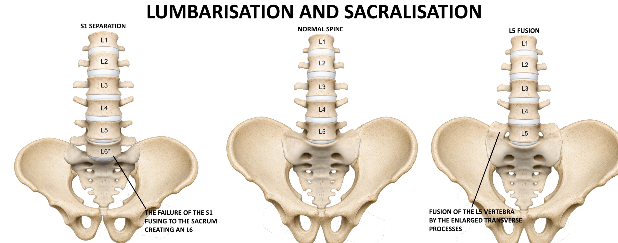
Variations in Lumbosacral Transitional Vertebrae (LSTV) occurs when there is an anatomical variation that can occur between a lumbar vertebra that has the characteristics of a sacral vertebra or vice versa. This is typically seen at the border of the last lumbar vertebra L5 and the first sacral vertebra S1.
In some individuals, L5 can develop additional features such as sacralisation and partially or fully fuse to the sacrum taking on the sacrum characteristics unilaterally or bilaterally.
Lumbarisation occurs when the S1 separates from the sacrum and becomes an additonal lumbar vertebra creating essentially an L6 and has all of the lumbar vertebral characteristics creating an additional mobile segment.
LSTV will have different characteristics but can include one of these characteristics: rudimentary joint presence (partial), lumbar sacral fusion with the sacrum (partial), and lumbar/sacral manifestations (absence).
The incidence of LSTV differs between populations with some studies indicating incidences in the range of 4-35% of a population meaning it can affect 6 out of 20 people and is quite common. However not all LSTV’s cause pain and most have no symptoms.
Implications of Anatomical Variations Lumbosacral transitional vertebrae may significantly change the anatomy and functioning of the spine due to the altered mechanics of the lumbosacral junction, and the potential for varying types of forces during movement at this joint, the surrounding structures (muscles, ligaments and intervertebral discs) could be subjected to an abnormal amount of stress.
The resulting instability may predispose some individuals to develop many spinal disorders such as:
degenerative disc disease, The discs above or below the transitional segment may experience increased wear and tear.
spondylolisthesis, occurs when one vertebra slips forward over the bone under neath it.
facet joint arthritis, The joints between the vertebrae can become irritated and develop arthritis due to abnormal stress.
Sacroiliac Joint Dysfunction, The abnormal connection can impact the function of the sacroiliac joints, which connect the sacrum to the pelvis.
Instability in cases of lumbarisation. the extra mobile segment can lead to instability.
Nerve Compression: In rare cases, the altered anatomy can contribute to compression of nerve roots exiting the spinal canal, leading to sciatica or radicular pain.
Anatomical variations can pose problems for doctors in both clinical evaluations and performing surgery. For example, the presence of LSTV might make it difficult to interpret diagnostic imaging, such as X-rays or MRIs, creating difficulty when diagnosing conditions that involve LSTV. Therefore, it is vital that healthcare professionals be familiar with these types of variations when creating treatment plans specifically for the patient and to ensure the vertebra disc count is also correct.
Understanding the anatomy of the lumbosacral region is important for recognising the significance of transitional vertebrae.
Variations among the lumbosacral vertebrae can help improve your understanding of spinal health as it relates to clinical practice.